Bild -
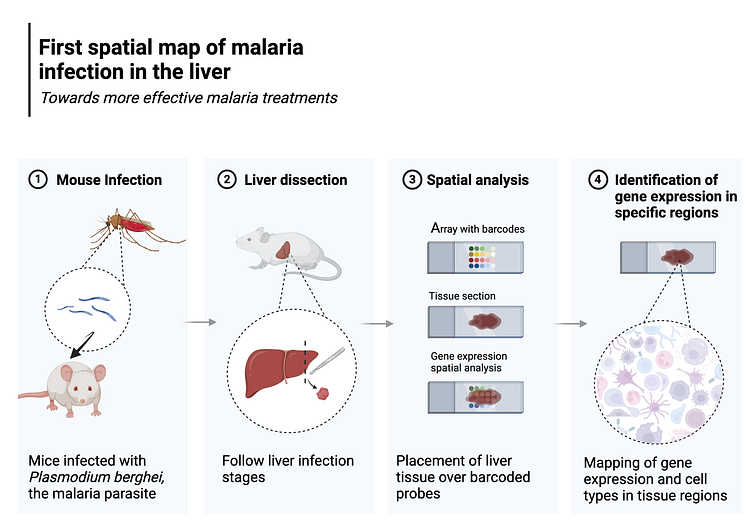
Study outline
1) Laboratory mice were infected with the rodent specific malaria parasite, Plasmodium berghei. The malaria infected livers were subsequently removed and snap-frozen at multiple time points, from early to late infection. 2) The snap-frozen tissues were cut into very thin, 10 micro-meter slices, and 3) placed onto a special microscopy slide covered in spatially barcoded molecular probes, which can capture RNA directly from the tissue above them. 4) Sequencing of the spatially barcoded RNA enables analyses of the expression of different genes. Thus, gene expression profiles were charted across the liver tissue samples to identify and compare gene expression across different tissue regions, such as parasite infected versus non-infected tissue areas (The figure was created with BioRender.com).
go to media item
- Licens:
- Medieanvändning
Innehållet får laddas ner, användas och delas i olika mediekanaler av t.ex. journalister, bloggare, krönikörer, opinionsbildare etc., i syftet att förmedla, redogöra för och kommentera ert pressmeddelande, inlägg eller information, så länge innehållet används oförändrat och i dess helhet. Upphovsmannen ska anges i den omfattning och på det sätt god sed kräver (vilket bl.a. innebär att fotografer till bilder nästan alltid måste anges).
- Upphovsrätt:
- Stockholms universitet
- Filformat:
- .png
- Storlek:
- 3000 x 2100, 644 KB