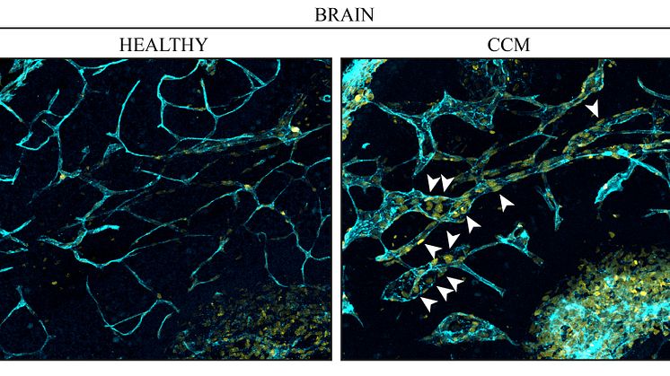
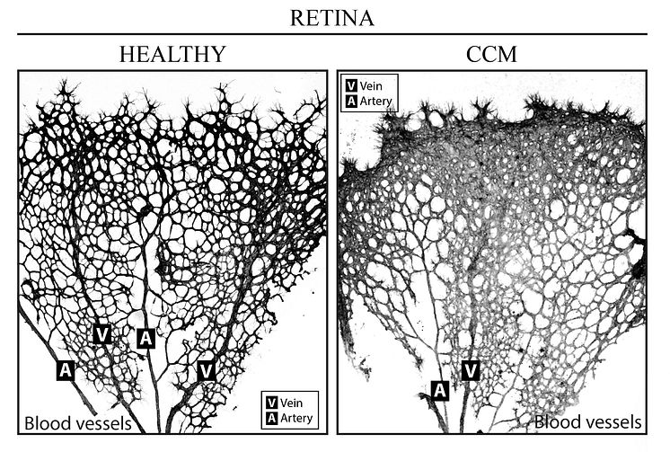
Skip to content
CCM retina

Image -

CCM retina

Veins (V) and arteries (A) in the retina. On the left a healthy retina is shown and on the right the same tissue from an individual with cavernoma.
Fabrizio Orsenigo
License:
Media Use
The content may be downloaded by journalists, bloggers, columnists, creators of public opinion, etc. It can be used and shared in different media channels to convey, narrate, and comment on your press releases, posts, or information, provided that the content is unmodified. The author or creator shall be attributed to the extent and in the manner required by good practice (this means, for example, that photographers should be attributed).
By:
Fabrizio Orsenigo
File format:
.jpg
Size:
2700 x 1819, 1.12 MB
Download

Contacts

Related content

  • Blood vessels in a brain. Left: endothelial cells (turquoise blue) from a healthy brain. Right: cavernoma leads to increased cell division in the endothelial cells (nucleus marked yellow) in the veins and causes malformations. Photo:Fabrizio Orsenigo

    Venous origin of brain blood-vessel malformations

    In the condition known as cavernoma, lesions arise in a cluster of blood vessels in the brain, spinal cord or retina. Researchers from Uppsala University can now show, at molecular level, that these changes originate in vein cells. This new knowledge of the condition creates potential for developing better therapies for patients. The study has been published in the journal eLife.