Image -
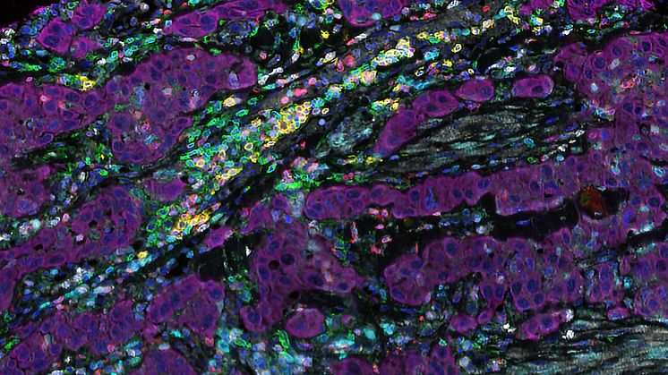
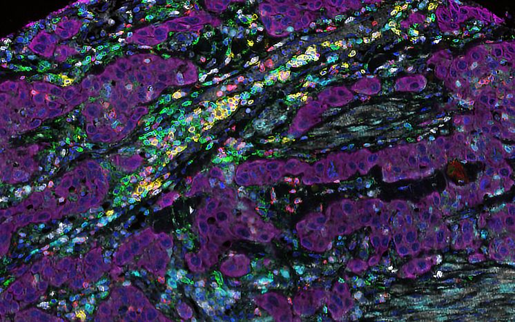
Human tumour tissue
Microscopic image of human tumour tissue (coloured magenta) and various immune cells: T cells (green and red), B cells (yellow) and NK (natural killer) cells (white). Activated T and B cells are depicted in cornflower blue, while the nuclei of all the cells are shown in the darker blue shade.
Iliana Kyriaki Kerzeli
- License:
- Media Use
The content may be downloaded by journalists, bloggers, columnists, creators of public opinion, etc. It can be used and shared in different media channels to convey, narrate, and comment on your press releases, posts, or information, provided that the content is unmodified. The author or creator shall be attributed to the extent and in the manner required by good practice (this means, for example, that photographers should be attributed).
- By:
- Iliana Kyriaki Kerzeli
- Copyright:
- Iliana Kyriaki Kerzeli
- File format:
- .tiff
- Size:
- 1361 x 852, 2.54 MB