Image -
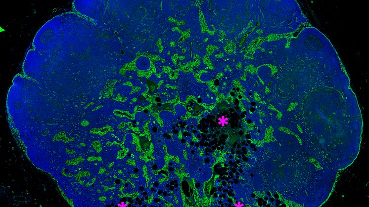
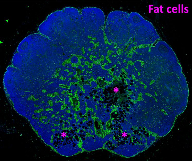
Lymph node with early stage lipomatosis
Immunofluorescence staining of a human lymph node (LN) with early stage lipomatosis (fat) in the medullary area of the LN. Adipocytes (fat cells shown as black, round cavities) are marked by asteriks' in magenta and their presence is associated with loss of medullary lymphatic sinuses (green).
Tove Bekkhus
- License:
- Media Use
The content may be downloaded by journalists, bloggers, columnists, creators of public opinion, etc. It can be used and shared in different media channels to convey, narrate, and comment on your press releases, posts, or information, provided that the content is unmodified. The author or creator shall be attributed to the extent and in the manner required by good practice (this means, for example, that photographers should be attributed).
- By:
- Tove Bekkhus
- File format:
- .jpg
- Size:
- 1947 x 1630, 4.33 MB