Pressmeddelande —
Bone regeneration after procedure for preservation of alveolar
Bone regeneration after procedure for preservation of alveolar ridge with Bond Bone
Dr. Kamen Kotsilkov (senior assistant at the Parodontology Department “ Faculty of Dental Medicine”, Medical University Sofia), Dr. Ivaylo Alyakov- DMD
Introduction
Ridge augmentation technics are a matter of choice after tooth extraction in order to prevent the loss of bone as a result of atrophic changes that may occur in time.
We can say that for the current widely used alloplastic materials based on betatricalcium phosphate and hidroxylapatite the resorbtion time is 6 – 9 - 12 months. This fact needs to be taken in consideration, when placing an implant in an area with guided bone regeneration. The need of using a membrane to immobilize the bone graft and covering it with flap additionally increases the complexity of the procedure.
In this case study we are going to demonstrate the capabilities of biphasic calcium sulphate (Bond Bone) as a bone graft material that provides predictable results and bone regeneration on the 4th month after the usage.
AIM
To present the histopathology result of bone regeneration in a clinical case of conventional implant placing after a procedure for saving the alveolar ridge using Bond Bone.
METHODS AND MATERIALS
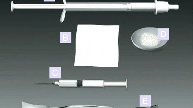
A 55 years old patient with present bridge constructions - #17_16_15_#14; #23_24_#25_#26; #34_35_36_#37; #44_45_46_#47 placed 5 years ago. The complaints of the patient are connected with recurring periodontic symptoms from tooth #14 (median bridge carrier from the bridge construction - #17_16_15_#14. After the removal of the bridge construction, a 2nd degree of mobility is determined as well as a periapical lesion. After an atraumatic extraction, the socket is filled in with Bond Bone without having to cover the graft with a membrane. Free gingival autotransplant from the neighboring toothless area is mobilized. The gingiva of the extraction wound is fixed with a modified vertical mattress suture stitch and the gingiva graft is fixated with O-stitches. After 4 months is performed a mucoperiostal flap with the purpose of placing dental implants on #14, #15 and #16. Bone biopsy is taken with a trephine borer from the places of the future implants. Sample 1 is taken from the spot with Bond Bone and Sample 2 is from patient’s own bone from spot #15.
Results of the histopathology- “Fragment of spongios bone with fibrosing of the bone marrow spaces”- Military Medical Academy, department “Patomorfology, autopsy and biopsy diagnostic”
The results of the biopsy are available at the importers office:
“Labtechnology” Ltd.
“Vranya” 82 str. Tel: 02/8320888
CONCLUSION
The presented clinical case shows that Bond Bone can be used with predictable success in cases where the bone volume of the alveoli ridge needs to be preserved. The advantages of the material are that there is no need to use an immobilizing membrane, as well as the quick hardening of the material in the presence of oral liquids. Having in mind that so far there isn’t a similar bone graft material with histomorphology on the 4th month close to natural bone, the product will be subject of additional research.
Pic.1 Ro condition before the extraction of #14 (14.06.2010)
Pic.2 Clinical condition before the extraction of #14 (16.06.2010)
Pic.3 extraction of 14 and immobilizing of free gingival autotransplant
Pic.4 after an atraumatic extraction the socket is filled with Bond Bone
Pic.5 Placement of the free gingival transplant of the toothless area
Pic.6 lagging of the extraction wound and a gingival transplant with O- stiches
Pic.7 removing of the thread (25.06.2010)
Pic.8 Clinical view before placement the dental implants (16.10.2010) 4 months later
Pic.9 Ro image of (CBCT) of the area with procedure of the volume preservation of the alveolar bridge with Bond Bone
Pic.10 planning of the implant positioning for the preparation of surgical model
Pic.11 Initial cut
Pic.12 Dissection of a flat in full width
Pic.13 Bone biopsy with trephine borer from the area of the future implants #14, #15 and #16
Pic.14 sample 1- histology
Pic.15 sample 2- control sample
Pic.16 the dental implants
Pic.17 reposition and fixation of the mucoperiostal flap with modified vertical mattress suture and O- stiches.
Pic.18 Ro condition after the surgical intervention
Relaterade länkar
Ämnen
- Utbildning
Kategorier
- alveolar
- augmenation
- regeneration
- bone
- tandimplantat
- calcium sulfate
- bondbone
- biphasic
- bifasiskt
MIS "Make it Simple" Implantat, Biomaterial och protetik
www.mis-implants.se