Image -
Picture montage periodontitis
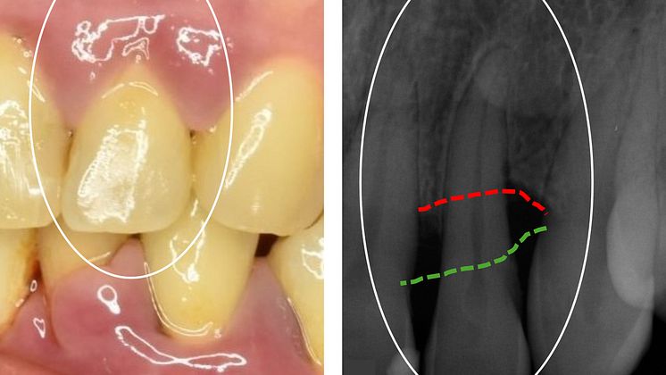
The image on the left shows a upper front tooth in an individual with periodontitis. On the right is an X-ray of the same tooth, revealing a significant reduction in the tooth's bone support. Normally, the jawbone level would be at the green line, but it has now receded to the red line. Consequently, the tooth root has almost no attachment to the jaw.
Elin Kindstedt
- License:
- Media Use
The content may be downloaded by journalists, bloggers, columnists, creators of public opinion, etc. It can be used and shared in different media channels to convey, narrate, and comment on your press releases, posts, or information, provided that the content is unmodified. The author or creator shall be attributed to the extent and in the manner required by good practice (this means, for example, that photographers should be attributed).
- By:
- Elin Kindstedt
- Copyright:
- Fri för publicering i medier
- File format:
- .jpg
- Size:
- 2564 x 1442, 1.21 MB