Image —
Image 2
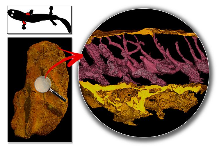
Top left: body outline of Acanthostega with the humeri represented in red. Bottom left: 3D model of a humerus of Acanthostega, generated from a synchrotron scan, indicating the position of the high-resolution model shown on the right. Right: 3D transverse section model through the outer layer of the bone showing blood vessel cavities (in pink) that provide clues about the metabolism.
Sophie Sanchez
- License:
- Creative Commons Attribution, no derivatives
With a Creative Commons license, you keep your copyright but allow people to copy and distribute your work provided they give you credit. You permit others to copy, distribute and transmit only unaltered copies of the work — not derivative works based on it.
- By:
- Sophie Sanchez
- File format:
- .jpg
- Size:
- 4355 x 3000, 5.59 MB