Image —
Inner ear
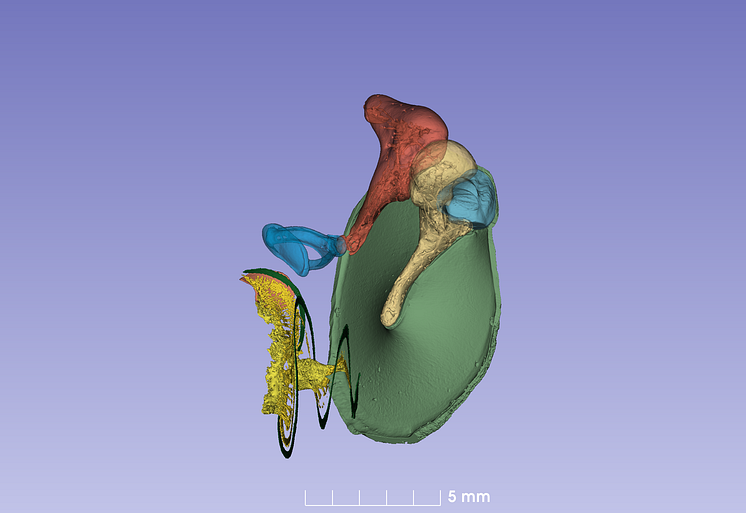
Three-dimensional image of the inner ear with all of its parts: The eardrum (green) and the auditory bones the malleus (light yellow), the incus (orange) and the stapes (blue). The green spiral at the bottom is the cochlea’s auditory membrane. There are hair cells on it that catch vibrations from the ear drum and the auditory bones. The yellow structure is the auditory nerve.
Springer Nature
- License:
- Media Use
The content may be downloaded by journalists, bloggers, columnists, creators of public opinion, etc. It can be used and shared in different media channels to convey, narrate, and comment on your press releases, posts, or information, provided that the content is unmodified. The author or creator shall be attributed to the extent and in the manner required by good practice (this means, for example, that photographers should be attributed).
- By:
- Springer Nature
- File format:
- .tiff
- Size:
- 2662 x 1830, 18.7 MB