Image —
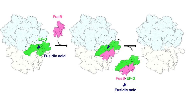
Schematic figure of the fusidic acid resistance “stop-motion” movie
Schematic figure of the fusidic acid resistance “stop-motion” movie. On the left, the protein EF-G (green) is locked to the ribosome (light blue and white) by the antibiotic fusidic acid (dark blue). In the middle, FusB (pink) binds and breaks EF-G off the ribosome, which releases fusidic acid. On the right, the rescued ribosome is ready to resume protein synthesis.
Maria Selmer
- License:
- Media Use
The content may be downloaded by journalists, bloggers, columnists, creators of public opinion, etc. It can be used and shared in different media channels to convey, narrate, and comment on your press releases, posts, or information, provided that the content is unmodified. The author or creator shall be attributed to the extent and in the manner required by good practice (this means, for example, that photographers should be attributed).
- By:
- Maria Selmer
- File format:
- .jpg
- Size:
- 1920 x 1080, 258 KB