Image —
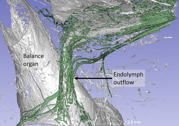
The human inner ear
A synchrotron X-ray of the balance organ of the human inner ear shows a kidney-shaped canal with a diameter of just approx. 0.5 mm. The inner ear has been reconstructed three-dimensionally in a computer program, and the surrounding bone has been made transparent. The green-coloured vessels surrounding the canal are thought to absorb and clean the fluid in the inner ear. It is believed that disruption of this function may cause Ménière's disease.
Uppsala University
- License:
- Media Use
The content may be downloaded by journalists, bloggers, columnists, creators of public opinion, etc. It can be used and shared in different media channels to convey, narrate, and comment on your press releases, posts, or information, provided that the content is unmodified. The author or creator shall be attributed to the extent and in the manner required by good practice (this means, for example, that photographers should be attributed).
- By:
- Uppsala University
- File format:
- .jpg
- Size:
- 2060 x 1443, 787 KB