Press release —
Advanced X-ray technology tells us more about Ménière's disease
The organ of balance in the inner ear is surrounded by the hardest bone in the body. Using synchrotron X-rays, researchers at Uppsala University have discovered a drainage system that may be assumed to play a major role in the onset of Ménière's disease, a common and troublesome disorder. These results are published in the journal Scientific Reports.
Ménière's disease is manifested in sudden onset of severe dizziness (vertigo) attacks, hearing impairment and tinnitus. Accumulation of excess fluid in the inner ear is thought to cause the disorder, from which approximately an estimated 30,000 people in Sweden suffer.
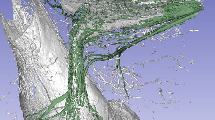
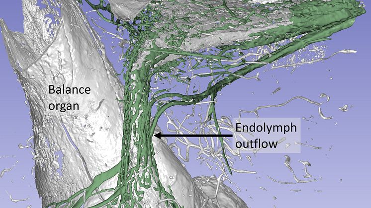
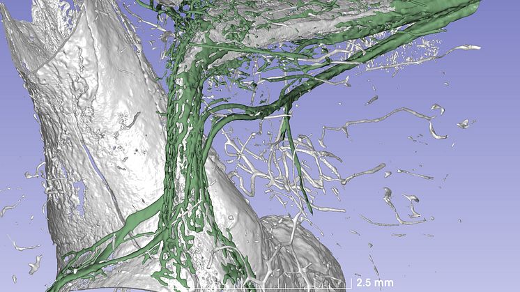
The researchers behind the new scientific article have investigated the organs in the human inner ear, which are very difficult to study. This part of the ear is enclosed by the body’s hardest bone. Using synchrotron X-ray imaging, an advanced and powerful form of computer tomography (CT), the scientists were able to study the organ of balance with its surrounding blood vessels. Since the technology generates energy too high for use on living humans, donors’ temporal bones were used.
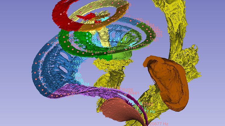
The images of the inner ear were reconstructed to make a three-dimensional model in the software, Inside the hard bone, the researchers discovered a drainage system that is thought to explain how the fluid in the inner ear is absorbed. This discovery may bring about an improved understanding of how and why Ménière's disease arises.
The synchrotron imaging investigation was carried out in Saskatoon, in the Canadian province of Saskatchewan. The study was conducted jointly with Dr Sumit Agrawal and Dr Hanif Ladak, who are researchers in London, Ontario (Canada).
For further information, please contact:
Charlotta Kämpfe Nordström, Physician at the Department of Surgical Sciences, Otorhinolaryngology and Head and Neck Surgery, Uppsala University, tel: + 46 70-3008951 email: charlotta.kampfe.nordstrom@surgsci.uu.se
Helge Rask-Andersen, Senior Professor at the Department of Surgical Sciences, Otorhinolaryngology and Head and Neck Surgery, Uppsala University tel: + 46 70-611 02 67, email: helge.rask-andersen@surgsci.uu.se
Nordström et al. (2020) A Micro-CT and Synchrotron Imaging Study of the Human Endolymphatic Duct with Special Reference to Endolymph Outflow and Meniere’s Disease, Scientific Reports, DOI:10.1038/s41598-020-65110-0
The study is also part of The Human Vestibular Aqueduct, Endolymphatic Duct and Sac: A Morphological Study Using Micro-CT, Super Resolution Immunohisto¬chemistry and Synchrotron Phase Contrast Imaging, a thesis publicly defended by Charlotta Kämpfe Nordström, an otologist at Uppsala University’s Department of Surgical Sciences, on 7 May 2020.
Topics
Uppsala University -- quality, knowledge, and creativity since 1477
World-class research and outstanding education of global benefit to society, business, and culture.
Uppsala University is one of northern Europe's highest ranked academic institutions. www.uu.se