News —
Clinical Case Studies: Axillary Breast Tissue (Polymastia) is common – even in men - you are not alone
Axillary breast tissue, also called polymastia or supernumerary breasts, is a common condition where extra breast tissue forms alongside the usual breasts. This extra tissue typically becomes noticeable during puberty.
It usually appears along the milk line, which runs from the chest to the groin. However, it can also occur in unusual areas, such as the face, back, and thighs.
While it doesn't function like normal breast tissue, it can sometimes develop lumps or, in rare cases, become cancerous.
Axillary breast tissue is not unusual; it can occur even in men. Here are its symptoms.
About 2% to 6% of women and 1% to 3% of men have axillary breast tissue. Some of them even have more than one area of extra tissue.
Common axillary breast tissue symptoms include swelling and tenderness in the area, thickening under the arms, limited shoulder movement, and irritation from clothing. These symptoms often worsen during puberty and pregnancy, when breast tissue naturally develops.
What does axillary breast tissue look like?


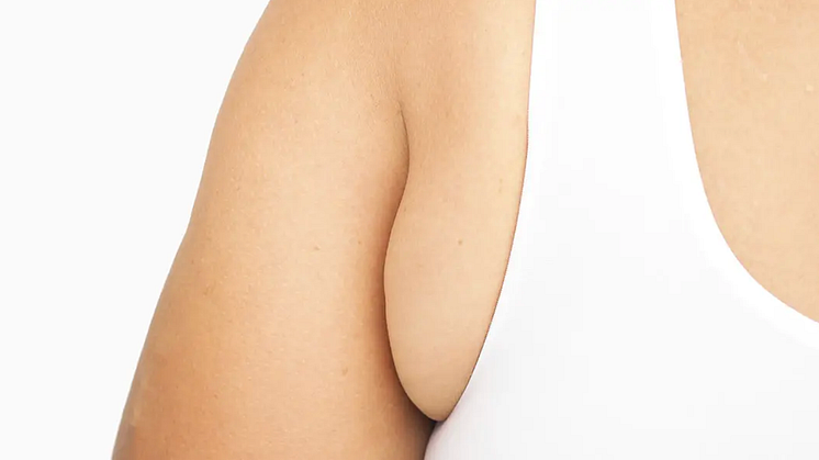
Axillary breast tissue appears as a lump, usually in the armpit area. This tissue can be noticeable when your arms are down and feels thicker than your usual fat pocket. Although it is generally harmless, it can sometimes feel tender or swollen during hormonal changes, which can cause cosmetic concerns.
Some axillary breast tissue can look like a breast, with an areola and nipple. How it looks and feels can vary based on its characteristics.
Grades of axillary breasts
According to Kajava, there are EIGHT grades of this condition. Grade IV is the most common.
- Grade I: A complete breast with a nipple, areola, and glandular tissue.
- Grade II: Glandular tissue and a nipple without an areola.
- Grade III: Glandular tissue and an areola without a nipple.
- Grade IV: Glandular tissue only.
- Grade V: The nipple and areola without glandular tissue.
- Grade VI: Only the nipple.
- Grade VII: Only the areola.
- Grade VIII: Only hair.
Case Study 1: 50-Year-Old Woman with Invasive Axillary Breast Cancer
Patient Description:
A 50-year-old woman developed invasive axillary breast cancer even though she had regular mammograms.
Diagnosis:
Doctors found a 2-cm tumour 4 cm below the left breast fold during a physical exam, and it had spread to the chest wall. A biopsy confirmed it was invasive stage IIIB ductal breast cancer.
The authors recommend diagnosing axillary breast cancer through physical exams and ultrasound. Removing the axillary breast may be the best treatment option if it shows symptoms.
Treatment Plan:
The patient first underwent chemotherapy before surgery, followed by more chemotherapy and radiation therapy afterwards.
This case shows that axillary mammary glands often go unnoticed in screening for breast cancer. Health professionals frequently use clinical exams and ultrasounds to diagnose axillary breast cancer.
For women with family history of breast cancer, preventive removal of the axillary breast is usually considered the best treatment.
Case Study 2: Treatment of Axillary Breast Tissue in female Patients of varied ages
Patient Overview:
Over 8 years, the researchers treated 29 female patients with axillary breast tissue, ranging from 19 to 54 years with the mean age being 28.8 years.16 had it on one side, and 13 on both sides of the armpit. Of these, 14 of them had breast hypertrophy.
Treatment Plans:
21 patients had surgery to only remove the breast tissue, while 5 only had liposuction, and 3 of them received both axillary breast tissue removal surgeries AND liposuction.
Postoperative Results:
All treatments — axillary breast tissue removal surgeries, liposuction, or both — produced good results.
Case Study 3: Large Axillary Breast Tissue in a 72-Year-Old Man
Patient Description:
A 72-year-old man presented with a swelling in his left armpit, which began about 50 years ago.
The swelling then was initially painless and measured about the size of a regular lemon. Throughout the decades, his axillary breast tissue has grown significantly bigger which extended beyond the armpit area, inducing discomfort and limiting his arm motion range.
Diagnosis:
A physical exam revealed a large, stalk-like mass in the left armpit, measuring 25 x 15 x 10 cm, with a smooth surface and no significant skin changes. Further tests that were conducted have confirmed glandular tissue; a biopsy showed a fibrous background with normal glandular cells and some fatty tissue.
Finding Kajava Grade I axillary breast tissue in older men is rare. It is crucial to consider this tissue when diagnosing long-term armpit swellings. The diagnosis included physical exams, imaging, and tissue analysis.
Treatment Plan:
Axillary breast tissue surgery
Postoperative Results:
Early detection and treatment of Kajava Grade I axillary breast tissue in older men with armpit swellings leads to better outcomes.
His surgery went smoothly and soon after, he felt physical relief from the symptoms. Axillary breast tissue surgery also reduced the risk of complications like cancer.

Case Study 4: A 71-Year-Old Woman with an Enlarged Extra Breast After Menopause
Patient Description:
A 71-year-old woman has an enlarged extra breast in her armpit that grew after she went through menopause.
Diagnosis:
Doctors confirmed the diagnosis through a tissue examination, which showed no signs of cancer. The results indicated growth of glandular tissue without an areola or nipple. This condition, known as Kajava Grade IV, is rarely reported in medical literature.
Treatment Plan:
The patient had surgery to improve her appearance and to lower the chances of cancer developing.
Case Study 5: A Mother in Her Mid-30s Concerned About Axillary Breast and Armpit Fat After Pregnancy
Patient Description:
A mother in her mid-30s sought help to address the lumps and armpit fat that she noticed during her pregnancy. The patient visited Dr Ivan Puah, an MOH-accredited liposuction doctor in Singapore with almost 20 years of body sculpting surgery experience, to find a permanent solution.
Diagnosis:
Dr Puah found axillary breast tissue AND fat in both her armpits.
Treatment Plan:
The combination of Axillary breast removal surgery and Modern Liposuction With MDC-Sculpt® Lipo Technique were recommended to remove the extra breast tissue and fat, as well as to tighten the skin around the armpit area.
Postoperative Results:
One week after the surgery, her incision sites healed well without any complications. She was happy with the results but worried about the scars. The doctor reassured her and suggested using scar gel, explaining that the scars would improve over time.

Is Axillary Breast Tissue Harmful?
A biopsy is usually recommended to check the tissue and rule out abnormalities. If surgery is needed to remove the extra breast tissue, the removed tissue will be evaluated for cancer or even carcinoma. Although the risk of cancer in this tissue is low (0.2% to 0.6%), it's highly recommended to send the excised axillary breast tissue for histological assessment.
